
Understanding Barrett's Esophagus
 Norman Barrett was born in Australia in 1903. He was educated in England, at Eton and Trinity College, Cambridge. Within 3 years of graduation, Barrett was elected to fellowship of the Royal College of Surgeons and had been awarded the postgraduate degree Master of Surgery. Supported by a Rockefeller Travelling Fellowship, Barrett studied in the United States in 1935 and 1936. Barrett went to America with the intent of studying upper gastrointestinal surgery, but he was urged by Donald C. Balfour at the Mayo clinic to enter into a newer field with more opportunities. That is when he settled on thoracic surgery.
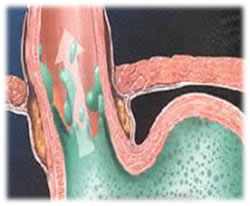
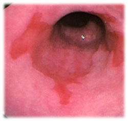
In 1950, Professor Norman Barrett noticed that, in certain patients, the distal esophagus had a lining that looked just like the lining of the stomach. That lining is called columnar epithelium. He originally believed that some people were born with this condition and described it “as tubular portion of the stomach being trapped in the chest”. Over time, other physicians argued that this stomach tissue was not stomach at all, but esophagus. It was suggested that these areas of stomach tissue on the lining of the esophagus be called “Barrett’s Ulcers”. Norman Barrett further described this condition as “lower esophagus lined by columnar epithelium”. Ever since, this disease described by Norman Barrett became known as “Barrett’s Esophagus”(BE).
So what is Barrett’s Esophagus? It is a condition referring to an abnormal change (metaplasia) in the cells of the distal portion of the esophagus. It is the replacement of a portion of the normal stratified squamous epithelium with columnar epithelium with goblet cells, which are usually found lower in the gastrointestinal tract. Barrett’s may also be referred to as intestinal metaplasia, or IM. It is further classified as low grade or high-grade dysplasia. Barrett’s is a precursor to esophageal cancer. It can can be readily seen on an esophagogastroduodenoscopy (EGD) or upper endoscopy, but many people who have reflux may have never had an EGD.
The following graphics depict an anatomic visualization and an endoscopic view.
Next Page: The Layers of the Esophageal Lining